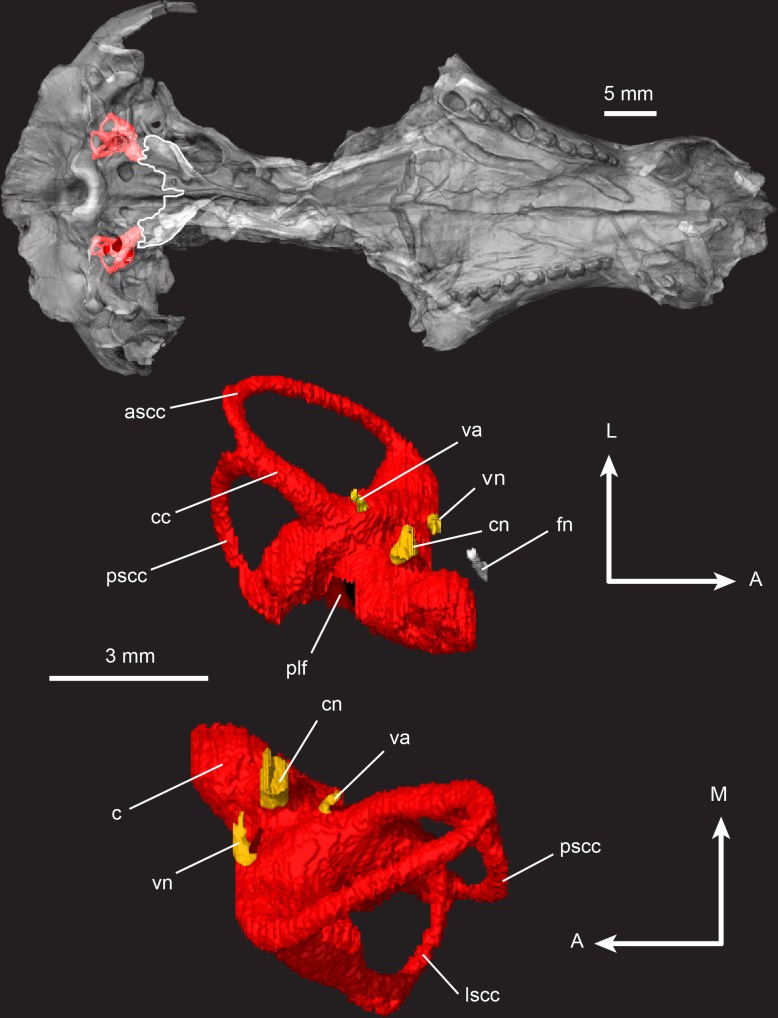
Fig 20. Pseudotherium argentinus, inner ear volume.
(Top) Ventral view of inner ear endocranial space in situ with a semitransparent isosurface model of skull. White tracings outline the parasphenoid alae and posterior border of basisphenoid. (Middle) Left inner ear volume in dorsal view. (Bottom) Left inner ear volume in ventral view. Abbreviations: ascc, anterior semicircular canal; c, cochlea; cc, common crus; cn, cochlear nerve (VIII); fn, facial nerve (VII); lscc, lateral semicircular canal; plf, perilymphatic foramen; pscc, posterior semicircular canal; va, vestibular aqueduct; vn, vestibular nerve (VIII). Arrow legend key: A = anterior, L = lateral, M = medial.

