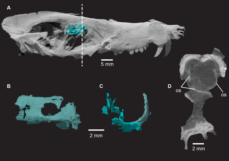
Fig 23.
Pseudotherium argentinus, orbitosphenoid (os) in situ (A), left lateral view (B), anterior view (C), and cross section (D). Left and right orbitosphenoids contact ventrally at midline. Left orbitosphenoid is less fractured than right orbitosphoid and shows distinct optic foramen. Cranium rendered semitransparent to illustrate relationship of orbitosphenoid to sphenorbital fissure and surrounding bony elements. Cross section illustrates the relatively dorsal position of the brain within the cranium.

