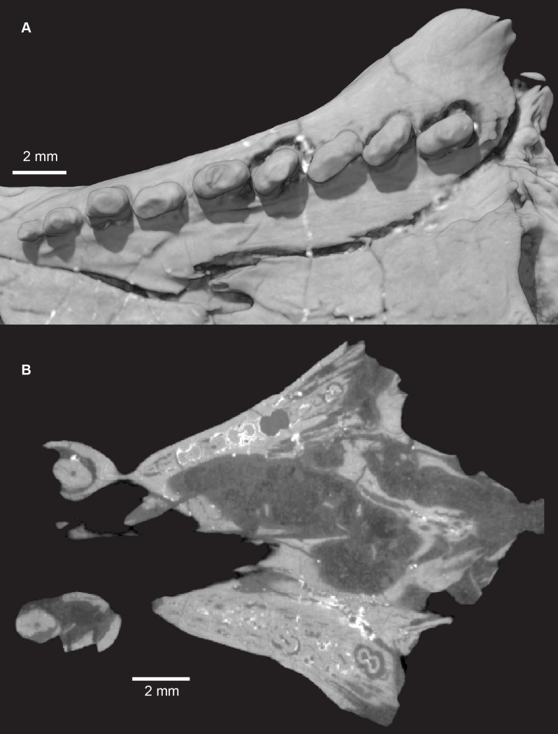
Fig 27. Pseudotherium argentinus, left postcanine tooth row.
(A) Occlusal view of left row in volume render. There are nine postcanines which increase in complexity from anterior to posterior, with the first postcanine being a single cusp and the penultimate postcanine having the most distinct cusps. Despite this trend, the postcanine cusps are small and blunt relative to postcanine cusps seen in other cynodonts. More posterior crowns (PC6-9) are mesiolingually in-turned. (B) Left maxillary tooth row in disto-occlusal view. Small accessory cusps are visible distobuccally on PC7 and PC8. (C) Horizontal section through the snout of PVSJ 882 illustrating constricted roots with a figure-eight cross section. The pulp cavity is compressed but never completely divided between root lobes. Nutrient canals run through each lobe of the root.

