Fig. 2.
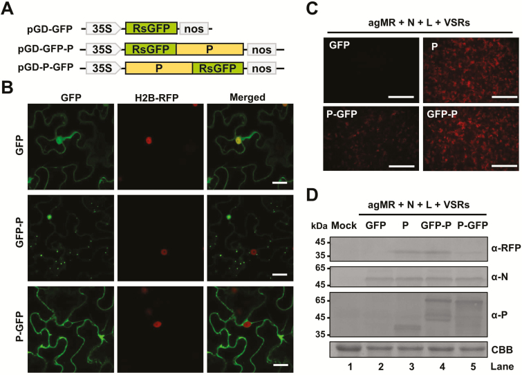
Subcellular localization and mobility of the BYSMV P protein. (A) Schematic diagram of pGD vectors for expression of free GFP, GFP-P, and P-GFP. (B) Confocal micrographs showing the subcellular distribution of free GFP, GFP-P, and P-GFP in epidermal cells of agro-infiltrated leaves of H2B-RFP transgenic N. benthamiana at 2 d post inoculation (dpi). Scale bars are 20 μm. (C) RFP foci at 6 dpi in leaves infiltrated with Agrobacterium engineered for expression of free GFP, P, GFP-P, or P-GFP in antigenomic-sense mini-replicon (agMR) combinations. Scale bars are 1 mm. (D) Western blotting analysis showing accumulation of RFP, and N and P proteins in infiltrated leaves with anti-RFP, anti-N, or anti-P polyclonal antibodies, respectively. Buffer-infiltrated leaves were used as a negative control (mock). Coomassie brilliant blue (CBB) staining was used for protein loading controls.

