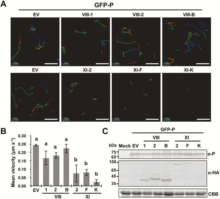
Fig. 5.
Effects of myosin tail overexpression on trafficking of BYSMV-P protein bodies in co-infiltrated N. benthamiana. (A) Representative confocal images of GFP-P bodies in epidermal cells overexpressing myosin tails VIII-1, VIII-2, VIII-B, XI-2, XI-F, and XI-K at 2 d post inoculation. Individual bodies were recorded in a time-series and are indicated by connecting lines. Different time intervals are represented by the different colors. (B) Mean velocities of the BYSMV-P bodies in the infiltrated leaves shown in (A). The mean velocities were calculated from the velocities of at least 90 bodies over 4 min. Data are means (±SE) of three independent experiments. Different letters indicate significant differences as determined by ANOVA followed by Turkey’s multiple comparison test (P<0.05). (C) Accumulation of GFP-P and myosin tails in those N. benthamiana leaves shown in (A). Anti-P, and anti-HA antibodies were used to detect the accumulation of GFP-P and HA-tagged myosin tails, respectively. Uninfiltrated N. benthamiana leaves were used as mock controls. Coomassie brilliant blue (CBB) staining was used for loading controls. EV, empty vector.

