. 2019 Jul 29;8:e48788. doi: 10.7554/eLife.48788

Figure 1. Lmx1b is required for the formation of ascending 5-HT axon projection pathways.

(A, B) Ascending 5-HT axonal projection system immunolabeled using an anti-RFP antibody to TdTomato in whole sagittal forebrain sections of 3 month old mice displayed by heatmap. Lmx1bcKO TdTomato+ axons were nearly absent in numerous brain regions (B) compared to controls (A) (n = 6, controls; n = 7, Lmx1bcKO adult mice). Right, schematics depicting 5-HT axon trajectories in Lmx1bcKO vs. control brains. Scale bars, 1000 µm. OB, olfactory bulb; ctx, cortex; cg, cingulum; cc, corpus callosum; hipp, hippocampus; spt, septum; hyp, hypothalamus; thal, thalamus; sc, superior colliculus; MFB, medial forebrain bundle; DRN, dorsal raphe nucleus. Schematic (right): ot, olfactory tract; cg/SCS, cingulum/supracallosal stria; fx, fornix; sm, stria medularis; fr, fasciculus retroflexus. (C) Confocal images of TdTomato+ axons in sagittal sections. Lmx1bcKO axons failed to fill cingulum bundles or innervate the hippocampus. Scale bars, 200 µm. cg, cingulum; cc, corpus callosum; LSM, lacunosum moleculare; DG, dentate gyrus; CA1 of hippocampus. (D) Coronal sections of cortex show near complete lack of Lmx1bcKO TdTomato+ axons Scale bars, 50 µm. (E) Coronal view of altered patterns of TdTomato+ axons in Lmx1bcKO thalamus. Arrowheads indicate areas devoid of axons in Lmx1bcKO thalamus. Numbers correspond to areas of axon clumping in Lmx1bcKO thalamus. See Figure 1—figure supplement 2 for high magnification images. Scale bars, 100 µm. PVT, paraventricular nucleus of the thalamus; 3V, third ventricle.

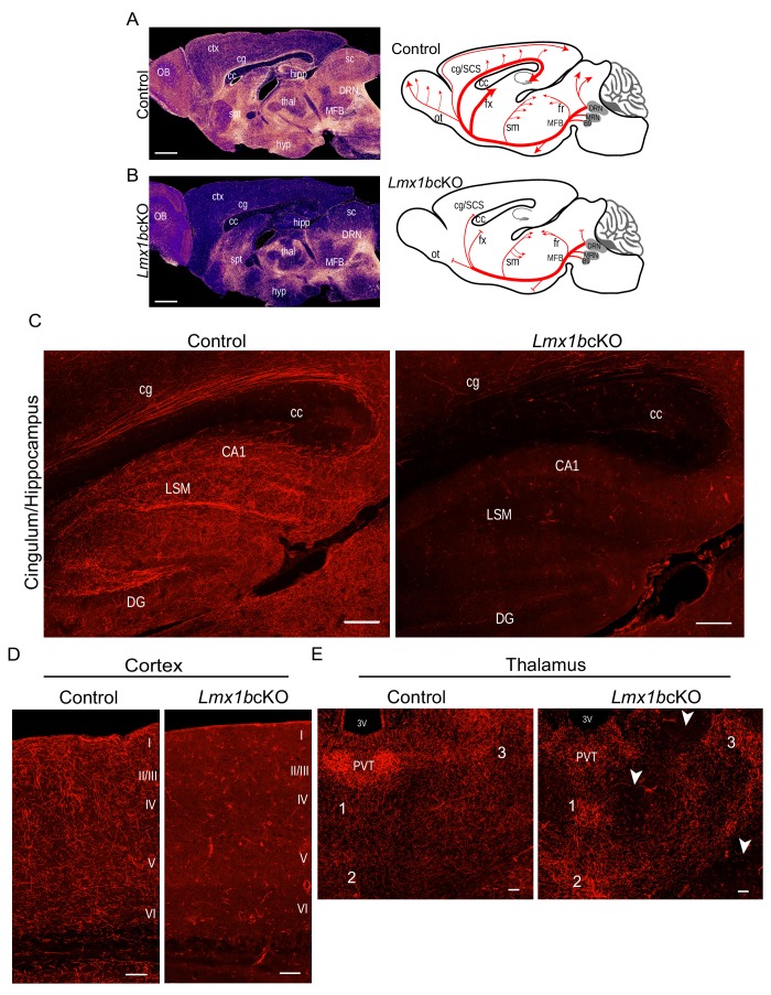
Figure 1.

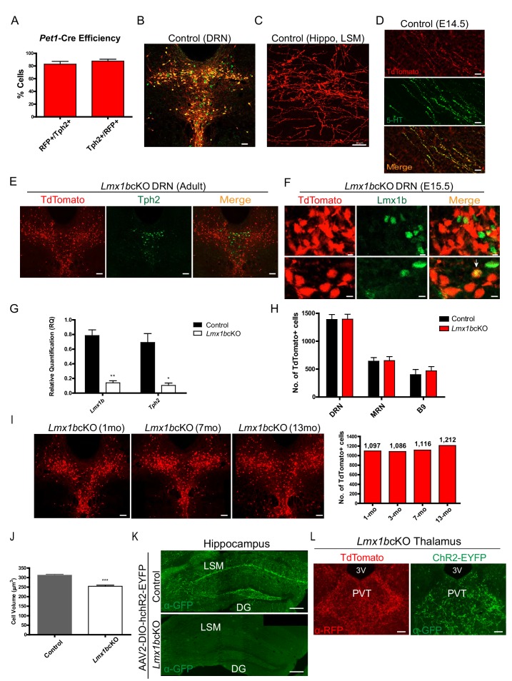
Figure 1—figure supplement 1. Surrogate marking of 5-HT cell bodies and axons and Lmx1b conditional targeting.

Figure 1—figure supplement 1.

(A) Pet1-Cre efficiency: 82% Tph2+ cells expressed TdTomato+ (RFP+/Tph2+) and 88% of TdTomato+ cells expressed Tph2 (Tph2+/RFP+) (n = 2 control mice). Data are represented as mean ± SEM. (B) TdTomato+ cells co-labeled with serotonergic marker Tph2 in the DRN. Immunofluorescence of Tph2 (green) and TdTomato (red). Scale bars, 100 µm. (C) TdTomato+ axons were found throughout the adult brain. Lacunosum moleculare layer (LSM) of hippocampus shown here. Scale bars, 10 µm. (D) Co-immunolabeled axons at embryonic day (E) 14 with anti-5-HT and anti-RFP antibodies. Scale bars, 20 µm. (E) A majority of TdTomato+ Lmx1bcKO mutant cells did not co-localize with Tph2 (n = 6, control; n = 7, Lmx1bcKO; 2.5–3.5 month old mice). Scale bars, 100 µm. (F) A majority of TdTomato+ Lmx1bcKO mutant cells did not co-localize with Lmx1b protein (E15.5 shown). A small number of cells were TdTomato+ and Lmx1b+ (bottom row, arrow). Scale bars, 5 µm. (G) RT-qPCR of Lmx1b and Tph2 in flow sorted YFP+ cells from control and Lmx1bcKO E17.5 embryos (n = 4, control; n = 4, Lmx1bcKO embryos). Unpaired t-test with Welch's correction, *p<0.05, **p<0.001, and ***p<0.0001. Data are represented as mean ± SEM. (H) Counts of TdTomato+ cells in Lmx1bcKO mice did not differ from controls. Every 4th section counted throughout DRN/MRN/B9 raphe nuclei (n = 3 mice/genotype). Data are represented as mean ± SEM. (I)  Lmx1bcKO cell bodies survive at least up to 13 months. Counts of TdTomato+ neurons in 6 matched sections for each animal (right) (n=1 animal per time point; 1mo, 3mo, 7mo, 13mo). Scale bars, 100µm. (I) Lmx1bcKO cell bodies survive at least up to 13 months. Counts of TdTomato+ neurons in 6 matched sections for each animal (right) (n=1 animal per time point; 1mo, 3mo, 7mo, 13mo). Scale bars, 100µm. (J) Volume of Lmx1bcKO cell bodies were significantly smaller than controls (n = 587 control cells; n = 442 Lmx1bcKO cells). Unpaired t-test with Welch's correction, *p<0.05, **p<0.001, and ***p<0.0001. Data are represented as mean ± SEM. (K, L) Expression of a YFP tagged channel rhodopsin by AAV2 viral injection to DRN/MRN of Lmx1bcKO and control mice. No YFP expression was found in Lmx1bcKO hippocampus (K) while YFP-labeled axons co-localized with TdTomato in the Lmx1bcKO thalamus (L). Scale bars, 200 µm (K), 50 µm (L).
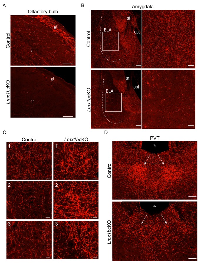
Figure 1—figure supplement 2. Lmx1b deficiency disrupts 5-HT axon patterns in the forebrain.

Figure 1—figure supplement 2.

(A) Sagittal sections of olfactory bulb show near complete lack of Lmx1bcKO TdTomato+ axons. Scale bars, 50 µm. gl, glomerular layer; gr, granule layer. (B) TdTomato+ axons severely reduced throughout Lmx1bcKO amygdala compared to controls (BLA, shown right). Scale bars, 100 µm. BLA, basolateral amygdala; st, stria terminalis; opt, optic tract. (C) High magnification showing areas of clumping (correlate to numbers in Figure 1E) found in Lmx1bcKO thalamus compared to controls. Scale bars, 20 µm. (D) Lack of TdTomato+ axons in the PVT of Lmx1bcKO mice. Scale bars, 50 µm. 3V, third ventricle; PVT, paraventricular nucleus of the thalamus.