. 2019 Jul 29;8:e48788. doi: 10.7554/eLife.48788

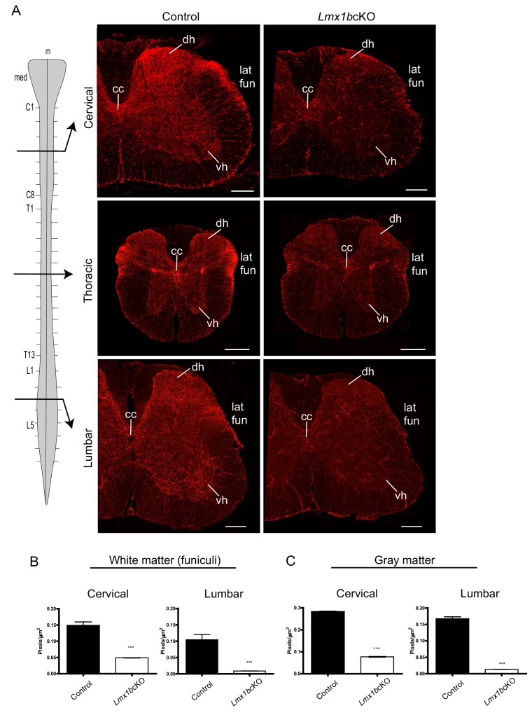
Figure 2. Lmx1b is required for the formation of descending 5-HT axon projection pathways.

(A) Coronal sections taken at cervical (C4), thoracic (T6), and lumbar (L3) levels of the spinal cord (diagram, left). Immunolabeling for TdTomato shows Lmx1bcKO axons were severely reduced at every level of the cord in both gray and white matter compared to controls. Scale bars, 200 µm. m, midline; med, medulla; cc, central canal; dh, dorsal horn; vh, ventral horn; lat fun, lateral funiculi. (B, C) Quantification of total TdTomato+ axons (pixels/µm2) in white (B) and gray (C) matter at cervical and lumbar levels (n = 3, control; n = 3 Lmx1bcKO mice). Two-way ANOVA with Welch’s correction, *p<0.05, **p<0.001, and ***p<0.0001. Data are represented as mean ± SEM.

Figure 2.

Figure 2—figure supplement 1. Conditional targeting of Lmx1b in the descending 5-HT projection pathway.

Figure 2—figure supplement 1.

(A) Pet1-Cre efficiency: 80% of Tph2+ cells expressed TdTomato+ (RFP+/Tph2+); 92% of TdTomato+ cells expressed Tph2 (Tph2+/RFP+) in medullary nuclei (n = 2 control mice). Data are represented as mean ± SEM. (B) TdTomato+ axon patterns in funiculi of the developing spinal cord aligned with 5- HT labeled axon patterns at E15.5 in controls (coronal view). Scale bars, 100 µm. (C) Comparable numbers of TdTomato+ medullary cells in Lmx1bcKO and control mice (n = 2 mice/genotype). (D) Few Lmx1bcKO TdTomato+ cell bodies expressed Tph2, a downstream target of Lmx1b. Scale bars, 100 µm.
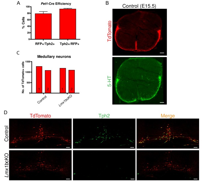
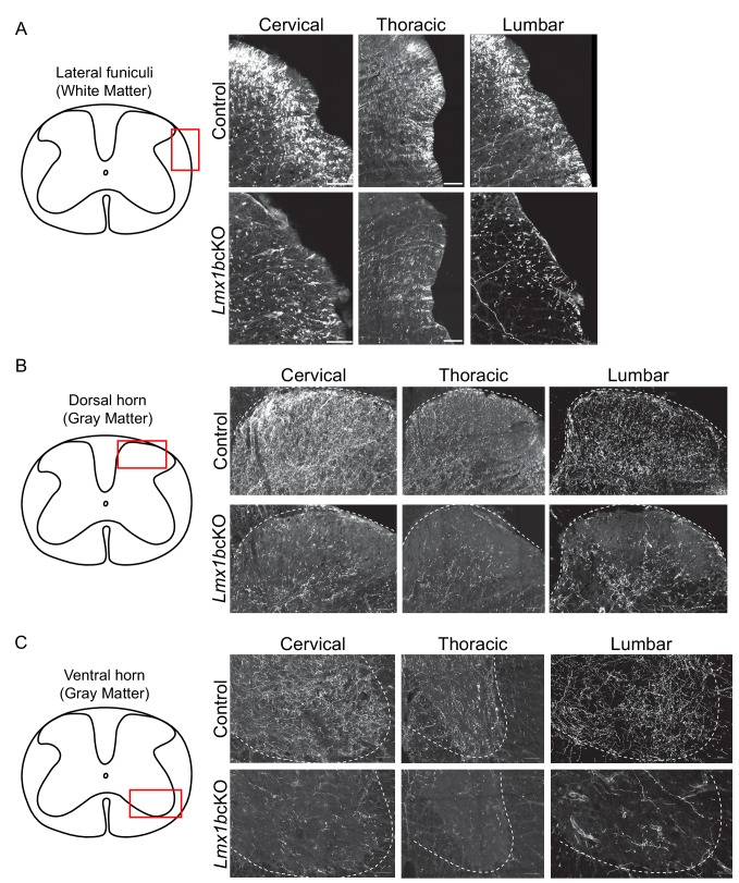
Figure 2—figure supplement 2. Progressive deficits of 5-HT axon fibers in Lmx1b deficient spinal cord white and gray matter.

Figure 2—figure supplement 2.

(A-C) Coronal sections immunolabeled using an anti-RFP antibody to TdTomato. Lmx1bcKO TdTomato+ axon deficits in spinal cord white matter funiculi (A), gray matter dorsal (B), and ventral (C) horns. Red box in diagram outlines area imaged (right). (n = 3, control; n = 3 Lmx1bcKO mice). Scale bars, 50 µm.