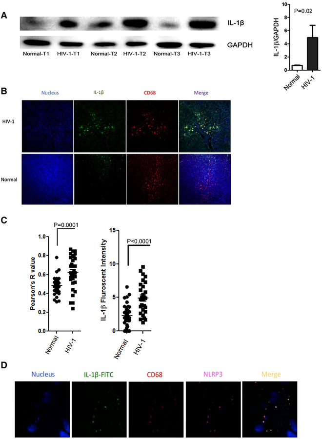
FIGURE 6. Immunoblot and Immunofluorescence staining reveals increased intrahepatic IL-1β expression in CD68+ macrophages in liver tissue from HIV-infected patients.
(A) IL-1β protein in human liver tissues was detected by Western blotting, GAPDH was used as internal control. Representative Western blot shown with densitometry from all specimens (P = 0.02) ((normal (n = 10) and HIV-1 monoinfected (n = 7)). (B) Confocal microscopic analysis of IL-1β and CD68 co-immunostaining in normal (n = 10) and HIV-1-infected liver tissues (n = 7). CD68 + liver macrophages (red), IL-1β (green) and nucleus (blue). (C) Colocalization analysis reveals increased co-localization of IL-1β and CD68 using image J in HIV-1 patients’ liver tissues compared to normal healthy livers (P = 0.0001). Increased IL-1β fluorescent intensity (= total intensity × Person’s R value) in HIV-1 liver tissue compared to normal livers (P < 0.0001). (D) Confocal microscopic analysis of IL-1β, NLRP3 and CD68 co-immunostaining in HIV-1-infected liver tissues. CD68 + liver macrophages (red), IL-1β (green), NLRP3 (magenta), and nucleus (blue). Panel A: Paired t test; Panel C: Welch’s test

