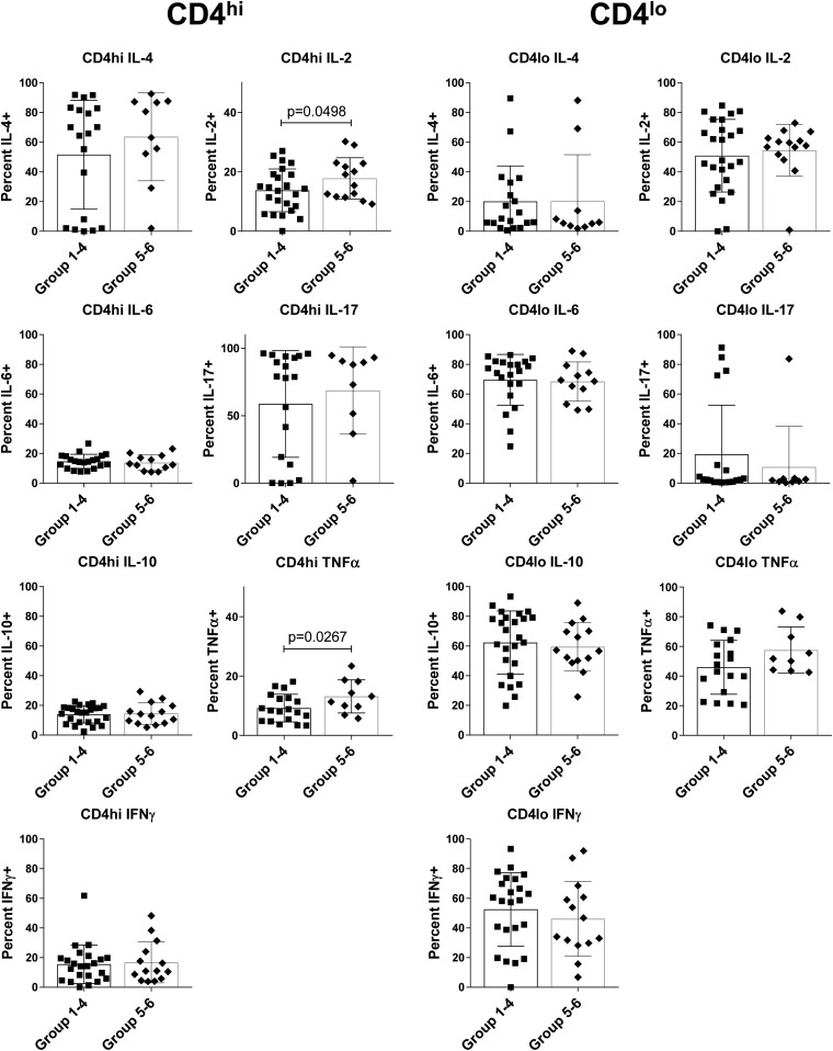
Figure 2.
Conventional CD4 T cells from stratified preT1D subjects demonstrate a difference in basal cytokine levels. PBMCs were stained for CD4, CD3, and CD40, as well as for intracellular cytokines. Cells were gated on FSC and SSC for live cells then on CD4lo and CD4hi on the basis of isotype control, and CD3 expression was confirmed. PreT1D subjects were stratified into groups as shown in Table 2, then subjects in groups 1 through 4 were compared with subjects in groups 5 and 6. Intracellular cytokine was measured by using the CD4−CD40− cells in each sample as a built-in negative control. Percentages of IL-2, IL-4, IL-6, IL-10, IL-17, IFNγ, and TNFα content in CD4hi (left panel) and CD4lo (right panel) are shown. Significant differences were calculated by one-tailed t test.

