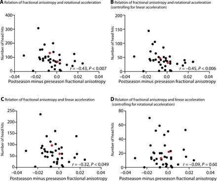
Fig. 3. Correlation between head hits with changes in FA.

In all plots, black circles indicate individuals without clinically diagnosed mTBI and red circles indicate the two individuals who suffered a concussion between the pre- and postseason MRI. (A) The scatter plot shows the relation between changes in FA in the right midbrain and the number of head impacts with rotational acceleration equal to or greater than 1 SD above the group mean. The direction of the relation indicates that more trauma is associated with greater reductions in the structural integrity of white matter. This relation remained when excluding the two participants who suffered a clinically defined concussion (r = −0.42, P < 0.012). (B) The relation between rotational acceleration and changes in FA holds when controlling for linear acceleration by restricting the analysis to hits that exceed the threshold for rotational acceleration but do not exceed the threshold for linear acceleration. This relation remained when excluding the two participants who suffered a clinically defined concussion (r = −0.44, P < 0.008). (C) The number of head hits with linear acceleration greater than 1 SD above the mean is negatively correlated with the change (postseason minus preseason) in FA in the midbrain. This relation, however, was not significant when excluding the two participants who suffered a clinically defined concussion (r = −0.32, P = 0.06). (D) The relation between linear acceleration and changes in FA goes away when controlling for rotational acceleration by restricting the analysis to hits that exceed the threshold for linear acceleration but do not exceed the threshold for rotational acceleration. This lack of a relation between linear acceleration and changes in DTI remained absent when excluding the two participants who suffered a clinically defined concussion (r = −0.11, P = 0.51).
