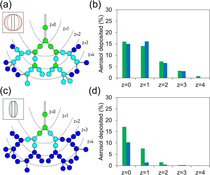
FIG. 4.
Exposure assays under different lung disease conditions. (a) Deposition pattern of the obstructivelike lung disease model. (b) Percentage of aerosol deposited in the channel (green) and bifurcation (blue) in each deposition zone for the obstructivelike breathing model. (c) Deposition pattern of the restrictivelike lung disease model. (d) Percentage of aerosol deposited in the channel (green) and bifurcation (blue) in each deposition zone for the restrictivelike breathing model.

