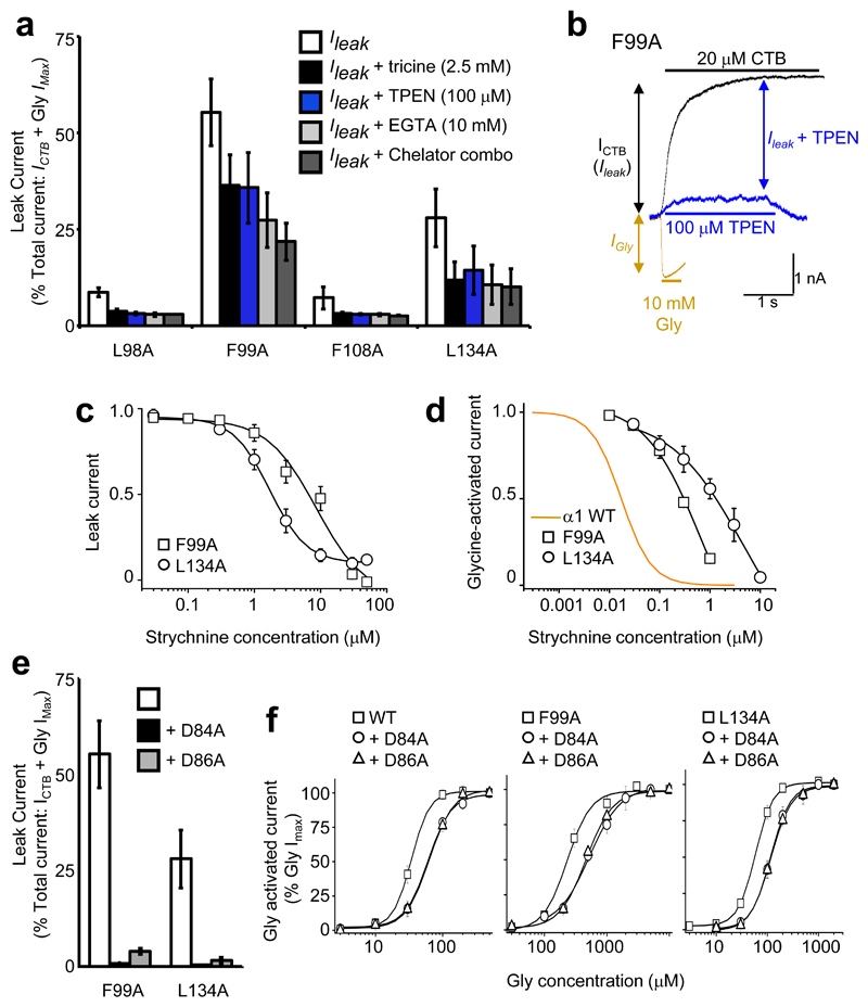
Figure 6. Spontaneous activation of alanine–substituted receptors.
(a) Persistent leak currents for alanine substituted GlyRs (= % CTB blockable current (ICTB)/(ICTB + Gly IMax)) in presence of individual and combined ion chelators. (b) Membrane currents for α1F99A ZAG in the presence of either glycine (orange), CTB (black) or TPEN (blue), overlaid to demonstrate their relative contributions to the total current (c) Strychnine concentration curves for inhibiting leak currents for α1F99A and α1L134A ZAGs (c) and EC50 glycine–activated current (d). Orange line indicates strychnine concentration inhibition curve for WT GlyRs, which are notably more sensitive as the agonist binding loops A and D are not perturbed. (e) Persistent leak current absent from receptors with alanine substitutions at Asp84 or Asp86 on α1F99A and α1L134A backgrounds. (f) Glycine sensitivity is modestly reduced in alanine–substituted Asp84 or Asp86 receptors, as compared to wild–type, α1F99A and α1L134A backgrounds. n = 3 –6.

