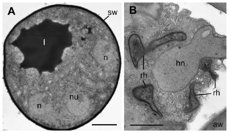
Figure 3.
Transmission electron micrographs of Apiochytrium granulosporum sp. nov. (X-124 CCPP ZIN RAS). (A) young sporangium with several nuclei and a big lipid globule, (B) host nucleus (hn) surrounded by rhizoids (rh). aw = algal cell wall; hn = host nucleus; l = lipid globule; n = nucleus; nu = nucleolus; rh = rhizoids; sw = sporangial wall. Scale bars: A—2 µm, B—1 µm.

