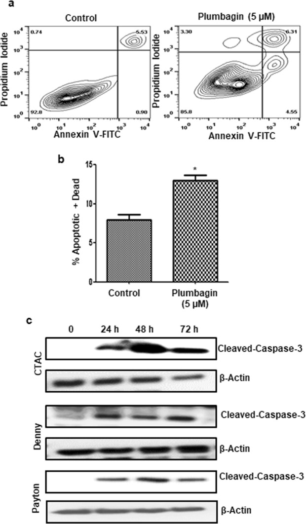
Figure 2.
Plumbagin induces apoptosis in canine cancer cells. (a) CTAC cells were treated with plumbagin (5 μM) for 24 h. Following treatment, the cells were stained with Annexin V-FITC (to detect expression of phosphatidylserine on the surface of the cell) and propidium iodide (as a live/dead cell indicator) and the cells were analyzed by analytical flow cytometry. (b) The bar chart shows average data from three independent replicates for the annexin V-FITC/propidium iodide assays. (*p = 0.006). (c) Three canine cancer cell lines CTAC, Denny and Payton were treated with plumbagin (5 μM) for the depicted time points. After treatment, the cells were lysed and expression of cleaved caspase 3 and β-actin (as a loading control) was determined by western blotting. The blots shown are representative of three independent replicates. Cropped blots showing cleaved caspase 3 and β- actin bands are shown. Full-length blots are provided in Supplementary Fig. 1.

