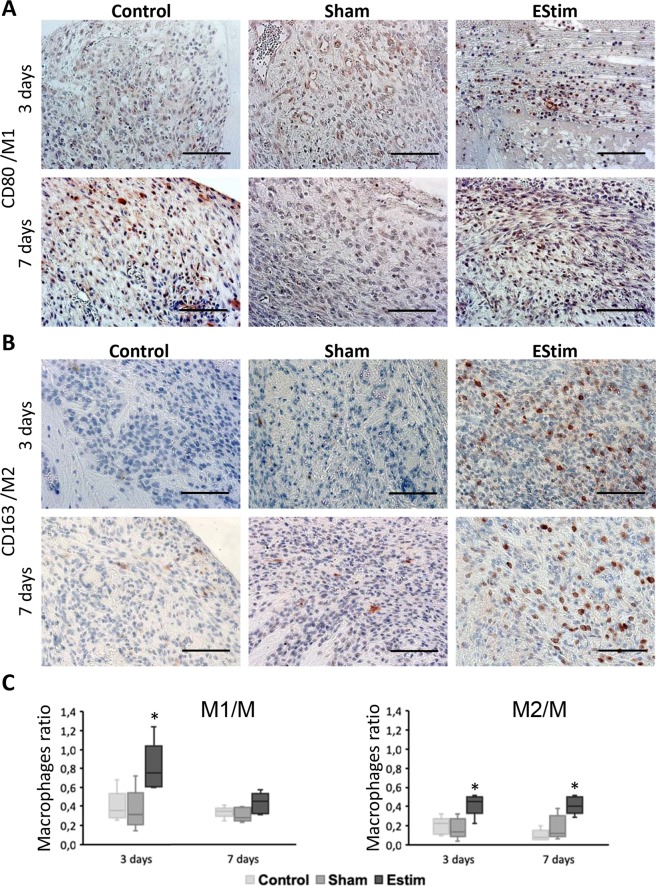
Figure 2.
Immunohistochemistry analysis of CD80 (marker for M1 macrophages) and CD163 (marker for M2 macrophages) in limb stumps at 3 and 7 days post-amputation. (A) Representative images of immunohistochemical staining showing higher incidence of M1 macrophages at day 3 in EStim treated tissues (Scale bar = 100 µm). (B) Representative images showing higher incidence of M2 macrophages at days 3 and 7 in EStim treated tissues (Scale bar = 100 µm). (C) Ratio of M1 and M2 macrophages (normalized to M macrophages) at the distal end of the stump for all groups tested at 3 and 7 days post-amputation; *p < 0.05.

