Figure 5.
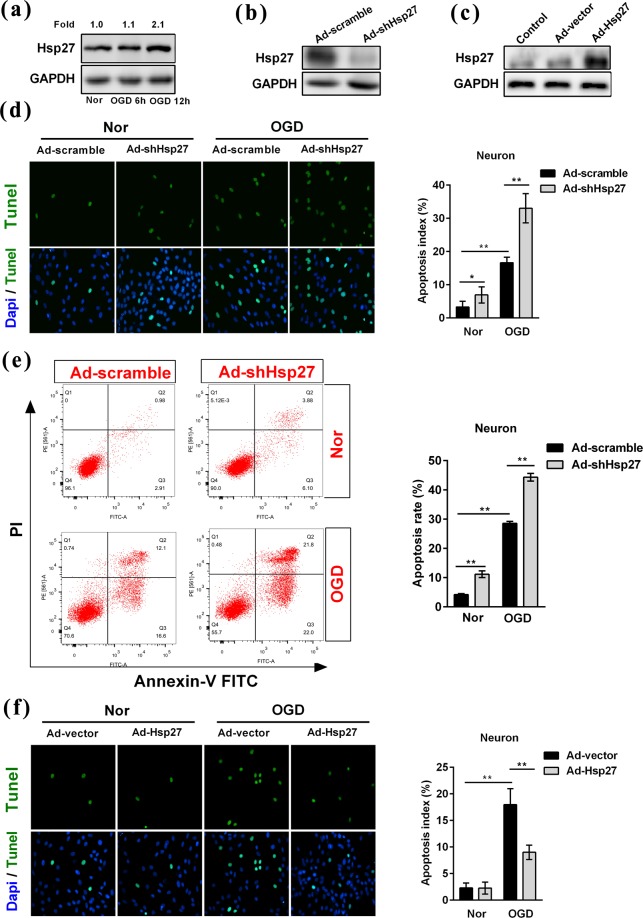
Hsp27 regulated the oxygen-glucose deprivation-induced apoptosis in primary rat neurons. (a) Primary neurons were treated by oxygen-glucose deprivation (OGD) at the indicated time points, and then Hsp27 protein was detected by western blotting in primary neurons. Primary neurons were transfected with the recombinant adenovirus carrying scramble RNA (Ad-scramble), short hairpin RNA-Hsp27 (Ad-shHsp27), empty vector (Ad-vector) or Hsp27 expression plasmid (Ad-Hsp27) for 48 h, and then neurons were cultured under normoxic conditions (Nor) or treated with OGD for another 12 h. Western blotting was performed to confirm the knock-down (b) and over-expression (c) effects of Hsp27 protein in normoxic neurons. (d,f) Tunel (Green) and Dapi (Blue) were assessed by immunofluorescence staining in transfected neurons under normoxic and OGD condition. The apoptosis index was calculated by dividing the number of apoptotic neurons (Green) by the total number of neurons (Blue). (e) Apoptosis was determined by Annexin-V FITC and PI staining and flow cytometry. Annexin-V (+)/PI (−) cells are early apoptotic cells and Annexin-V (+)/PI (+) cells are late apoptotic cells. The FACS analysis graphs are representative of three independent experiments. The rate of apoptosis was early apoptosis percentage plus late apoptosis percentage. Data were expressed as mean ± SD. *P < 0.05 versus the scramble- or vector-transfected cells under normoxia, **P < 0.01 versus the scramble- or vector-transfected cells under normoxia or the scramble- or vector-transfected cells treated by OGD.

