
Figure 1.
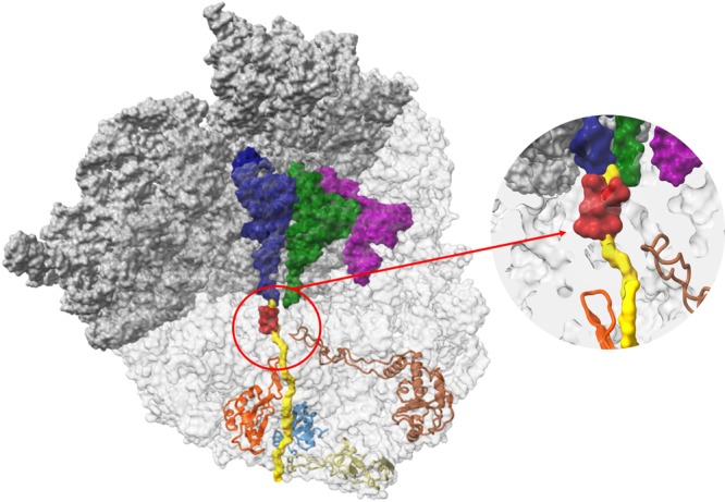
The ribosome nascent chain tunnel environment Left: The 70S S. aureus (SA_WT) ribosome where the large subunit is shown in light grey and the small subunit is shown in dark grey (PDBID 5TCU). The A-site, P-site and E-site docked tRNA molecules (from PDBID 5JTE) are shown in blue, green and magenta, respectively. The surface of a nascent chain within the tunnel is shown in yellow. Erythromycin surface is shown in red, uL4, uL22, uL23 and uL24 are shown in brown, orange, teal and khaki, respectively. Right: zoom into the ery binding site at the upper tunnel.