
Figure 3.
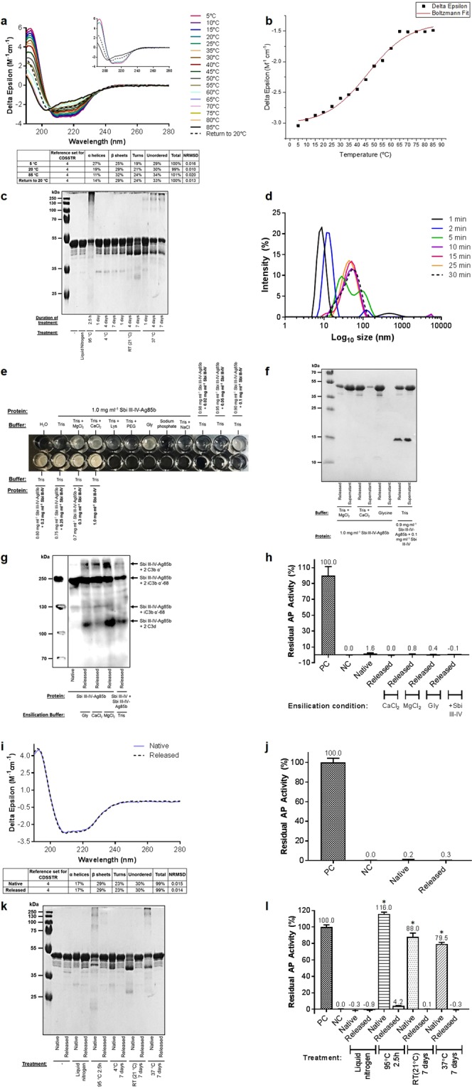
Ensilication protects Sbi III-IV-Ag85b against thermal denaturation. (a) CD spectra showing partially reversible thermal unfolding of Sbi III-IV-Ag85b. CD spectra at 5 °C, 20 °C, 85 °C and upon return to 20 °C are displayed in the inset figure. Deconvolution was performed using DichroWeb. (b) Boltzmann sigmoidal fit of CD thermal denaturation curve at 222 nm indicating a melting temperature of 46.3 °C. (c) Temperature-induced loss of Sbi III-IV-Ag85b structural integrity visualised by SDS-PAGE. (d) Time-course dynamic light scattering size distribution by intensity plot of Sbi III-IV-Ag85b ensilication in 50 mM Tris pH 7 showing a maximal particle size of 55 nm. (e) Sbi III-IV-Ag85b ensilication trials for visualisation of sol-gel precipitates showing that MgCl2, CaCl2, glycine and Sbi III-IV improve precipitate formation. Water and Sbi III-IV alone were included as negative and positive controls respectively. (f) SDS-PAGE analysis showing protein levels in the released solution and unensilicated supernatant following ensilication of Sbi III-IV-Ag85b under the four conditions depicted. See Supplementary Table S3 for protein concentrations. (g) Anti-Sbi western blot analysis of C3 activation in normal human serum following incubation with native or released Sbi III-IV-Ag85b. Higher molecular weight bands depicting opsonisation of native and released Sbi III-IV-Ag85b with C3 activation products are evident. All samples are at a concentration of 27 μM except for the released sample containing Sbi III-IV which is at 18 μM. (h) Alternative pathway activity analysis showing comparable complement depletion activity for native Sbi III-IV-Ag85b (4 µM) and Sbi III-IV-Ag85b (4 µM) released following ensilication under the conditions shown. Data are displayed as mean values (n = ≥ 2) ± standard deviation from the mean. A one-way ANOVA with Dunnett’s multiple comparisons analysis confirmed no significant differences in activity between native Sbi III-IV-Ag85b and the released Sbi III-IV-Ag85b samples (P > 0.05). (i) CD spectroscopy showing the near-identical secondary structures of native and released Sbi III-IV-Ag85b. Spectra are representative of 3 replicates. Deconvolution was performed using DichroWeb. (j) Alternative pathway assay depicting comparable functional activities for native Sbi III-IV-Ag85b (4 μM) and Sbi III-IV-Ag85b (4 µM) released subsequent to ensilication in 50 mM glycine pH 7.4. Data are displayed as mean values (n = ≥ 2) ± standard deviation from the mean. An unpaired t-test confirmed no significant difference in activity between native and released Sbi III-IV-Ag85b (P > 0.05). (k) SDS-PAGE profile illustrating thermal denaturation and aggregation in native but not released Sbi III-IV-Ag85b samples. (l) Alternative pathway activity analysis displaying retention of complement depletion activity in released but not in native thermally-treated Sbi III-IV-Ag85b (4 μM). Data are displayed as mean values (n = ≥ 2) ± standard deviation from the mean. A one-way ANOVA with Dunnett’s multiple comparisons analysis was used to analyse differences in activity compared to native untreated Sbi III-IV-Ag85b. *P < 0.01. DLS, CD and opsonisation analysis of thermally-treated native and released Sbi III-IV-Ag85b can be found in Supplementary Figs S4–S6, respectively. Full-length gel images of (c,f,k) are included in Supplementary Figs S17–S19 and full-length western blot image of (g) is shown in Supplementary Fig. S20.