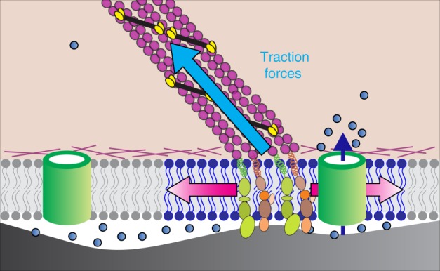
Fig. 7.

Working model of Piezo1 activation by traction forces. Traction forces (blue arrow) generated by Myosin II motors (yellow and black) along actin filaments (purple) tethered to integrin-based focal adhesion zones (green and tan) generate local increases in membrane tension (depicted by blue region of lipid bilayer and red arrows) that elicit Ca2+ flickers from nearby Piezo1 channels (green cylinder in the right part of the figure). Blue dots represent Ca2+ ions and dark blue arrow represents Ca2+ influx through Piezo1. Piezo1 channels far from force-producing adhesions are not activated (green cylinder in left of figure)
