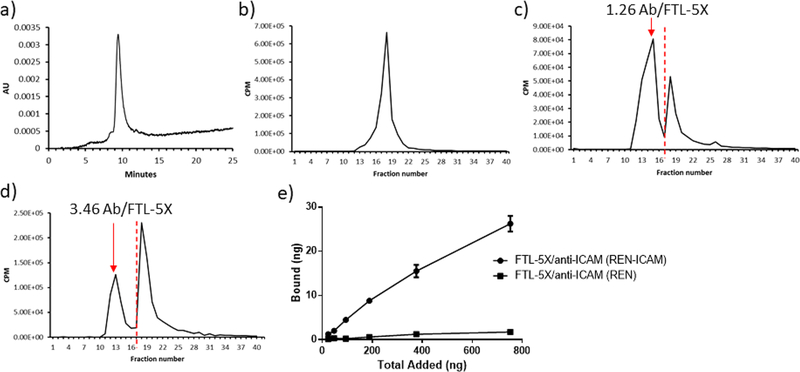
Figure 3.
Quantitative analysis of number of radiolabeled whole antibody conjugated to FTL-5X using HPLC. Antibody was radiolabeled with 125I, followed by conjugation to FTL-5X at different molar ratios. HPLC absorbance trace of (a) FTL-5X, and radiotraces of (b) anti-ICAM, (c) FTL-5X/ anti-ICAM (1 to 2 molar ratio), (d) FTL-5X/anti-ICAM (1 to 10 molar ratio). Red colored dashed line indicates the cutoff line used for calculation of areas under the curve (AUC) for conjugate vs free antibody peaks. (e) In vitro binding of 125I-labeled targeted FTL-5X to ICAM positive and negative REN cells. FTL-5X was 125I-labeled prior to antibody conjugation. Cells were grown to confluence and incubated with targeted FTL-5X nanocarriers for 1 h at 37 °C. Bound radiolabeled targeted nanocages were measured by gamma counter.

