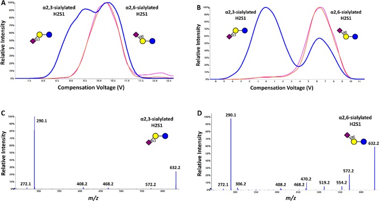
Figure 3.
Separation of deprotonated sialylated glycans H2S1 using DMS. The blue trace was obtained during the analysis of the mixture of the two isomers, while the red and pink traces were produced during the analysis of only the H2S1 isomer. While minimal separation is observed when the DMS is operated at SV = 4500 V using nitrogen alone as the carrier gas (A), the α2,3 and α2,6 sialic acid-linked isomers were fully separated when methanol was added to the carrier gas. Full scan MS/MS spectra (collision energy = 45 eV, lab frame for both spectra) obtained using the SV and CoV settings for full separation, show different fragment patterns for the α2,3 (C) and the α2,6 isomers (D). Note, the presence of a α2,6 isomer-specific 0,4A2-CO2 fragment at m/z 306 (D).

