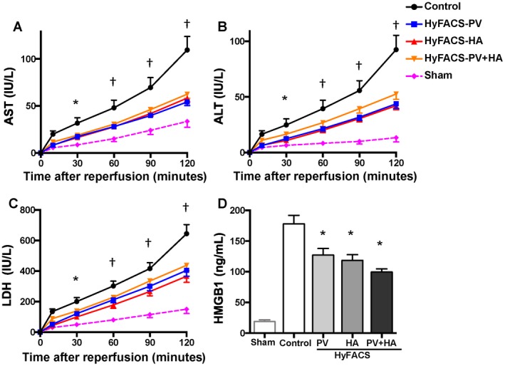
Figure 2.

Transaminase and HMGB1 release upon reperfusion. (A) AST, (B) ALT, and (C) LDH release into the perfusate, as indices for hepatocellular damage upon reperfusion. All differences among the groups were assessed via 2‐way repeated‐measures ANOVA (AST, P < 0.001; ALT, P < 0.01; LDH, P < 0.01). Time point assessments were performed by Bonferroni’s posttest (*P < 0.05; † P < 0.001; versus control group). Error bars are sometimes invisible due to small standard errors of the mean (n = 10 each). (D) HMGB1 release into the perfusate served as an index for comprehensive tissue damage after 2 hours of oxygenated reperfusion as well as for a hazardous proinflammatory signal thereafter. All data are presented as the mean ± standard error of the mean (n = 10 each). All differences among the groups were assessed via 1‐way ANOVA (P < 0.001) followed by Bonferroni’s posttest. *P < 0.05 versus other groups.
