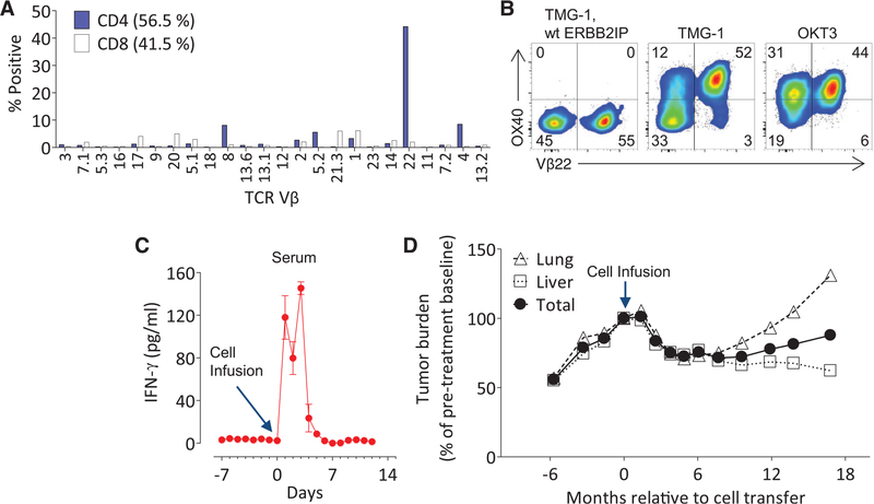
Fig. 2. Adoptive transfer ofTILcontainingERBB2IP mutation–reactive T cells.
(A) Flow-cytometric analysis of the TCR-Vβ repertoire of 3737-TIL, gated on live CD4+ or CD8+ T cells. (B) Patient 3737–TIL were cocultured with DCs transfected with TMG-1 or TMG-1 encoding the wt ERBB2IP reversion, and flow cytometry was used to assess OX40 and Vβ22 expression on CD4+ T cells at 24 hours post-stimulation. Plate-bound OKT3 stimulation was used as a positive control. (A) and (B) are representative of at least two independent experiments. (C) IFN-γ enzyme-linked immunosorbent assay on patient 3737 serum samples pre- and post-adoptive cell transfer of 3737-TIL. Error bars are SEM. (D) Tumor growth curves [Response Evaluation Criteria in Solid Tumors (RECIST), sum of maximum diameters] before and after infusion of 3737-TIL. Data are expressed as a change in percent from pretreatment baseline and stratified on lung, liver, and total tumors.

