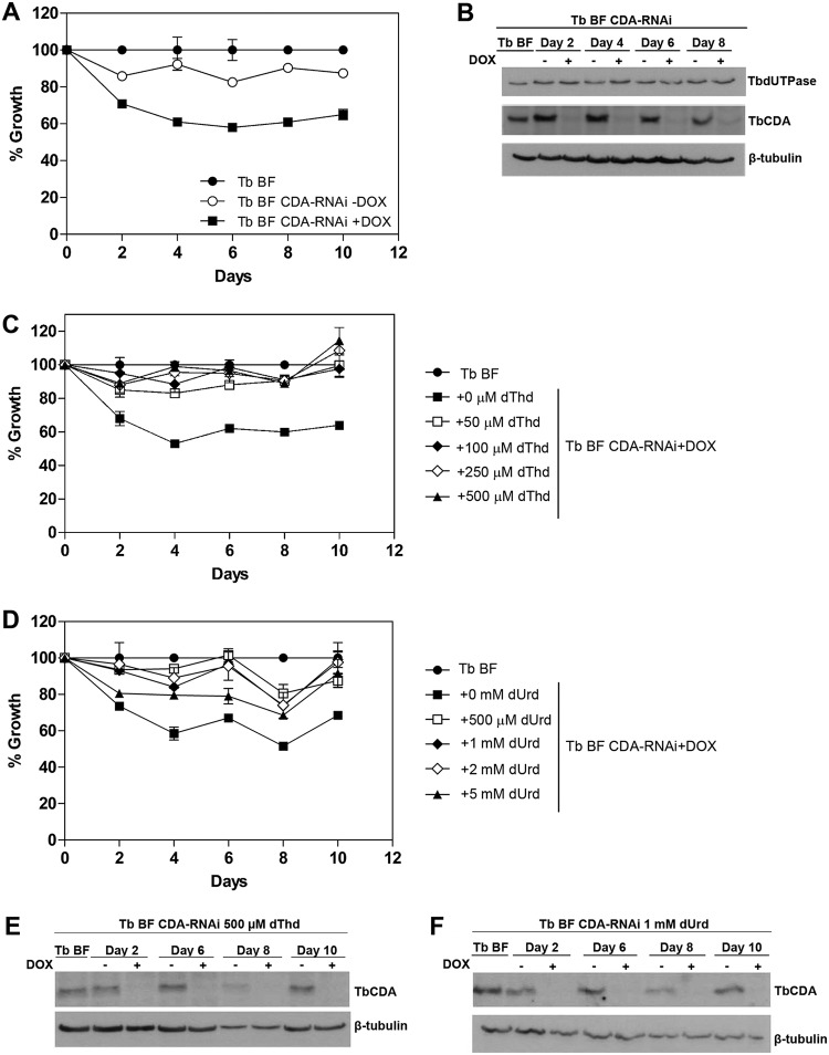
FIG 2.
(A) Growth curves of T. brucei BF and BF CDA-RNAi cell lines. DOX, doxycycline. (B) Western blot showing that TbCDA silencing was stable throughout the growth curve and that TbdUTPase levels remain unchanged after 10 days of TbCDA knockdown. (C and D) Growth curve of T. brucei BF and BF CDA-RNAi cell lines supplemented with different dThd (C) and dUrd (D) concentrations. Each point represents the mean from three biological replicates. Error bars represent standard deviations. (E and F) Western blot analysis demonstrating decreased TbCDA expression during supplementation with dThd and dUrd. Anti-TbCDA (1:500), anti-TbdUTPase (1:75,000), and anti-β-tubulin (1:5,000) antibodies were used. A total of 5 × 10⁶ parasites were loaded in each lane.

