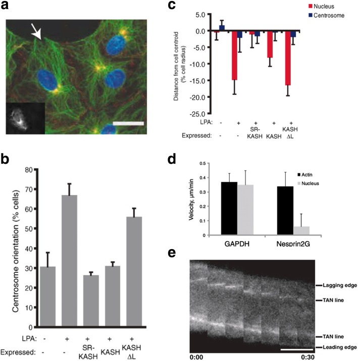
Fig. 2.
Cytokine-driven centrosome orientation requires nesprin-2G. NIH3T3 fibroblasts expressing dominant negative constructs (RFP-Spectrin Repeat-KASH, or RFP-SR-KASH, and RFP-KASH) targeting the LINC complex were stimulated with lysophosphatidic acid (LPA). a Epifluorescence image showing centrosome orientation of RFP-KASH-expressing cells (cell expressing RFP-KASH is indicated by arrow and in the inset). DNA (blue); centrosomes (yellow); microtubules (green); β-catenin for cell-cell adhesion (red). Scale bar = 15 μm. b Centrosome orientation by LPA treatment was impaired by dominant negative KASH. c Average centrosome and nucleus positions perpendicular to the wound (positive values are towards the leading edge; negative values are away). Nuclear movement by LPA was disabled by dominant negative KASH. d Nucleus velocity was decreased for nesprin2G-depleted cells. e Fluorescence kymograph of TAN lines in a nesprin2G-depleted nucleus during nuclear movement with time (hour:min). Scale bar = 5 μm. Adapted and reprinted with permission from [25]

