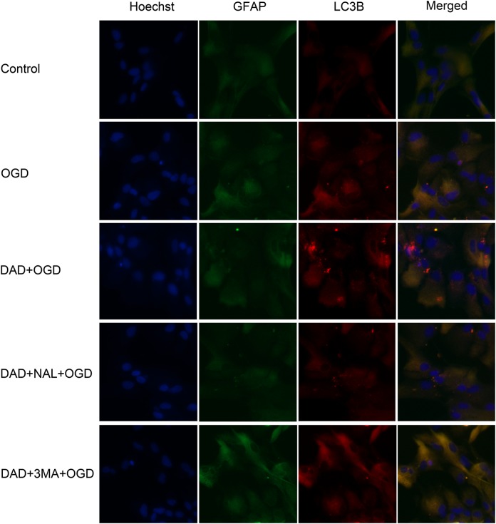
Fig. 2.
Representative images of LC3 fluorescence staining. Astrocytes were stained using GFAP antibody. Autophagic vacuoles were stained using LC3 antibody, and the cell nuclei were stained with Hoechst. Photomicrographs were taken under an epifluorescence microscope (magnification, x400). DAD, DADLE; NAL, naltrindole; 3-MA, 3-methyladenine.

