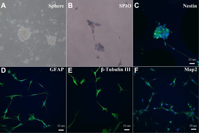
Figure 2.
iPS cells-induced NSCs, SPIOs labeling and identification. iPS cells-induced NSCs neurospheres were observed under microscope after passage (A); SPIOs-labeled NSCs had blue particles in the cytoplasm revealed by Prussian blue staining (B); SPIOs-labeled NSCs were identified with immunofluorescence staining for Nestin (C), GFAP (D), β-Tubulin III (E), and Map2 (F).

