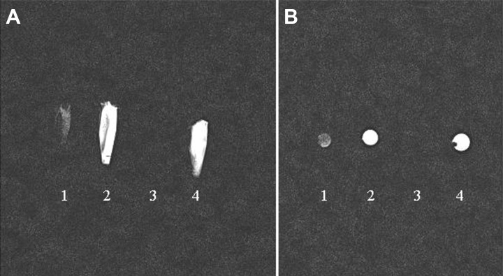
Figure 3.
In vitro MRI scanning of iPS cells-derived NSCs and solutions. The data showed that SPIOs-labeled NSCs (1) presented with hypointense signals compared with unlabeled cells (2) and cell medium (4). However, the SPIOs solution could not be visualized due to high magnetic field effect (3). (A: sagittal view; B: axial view).

