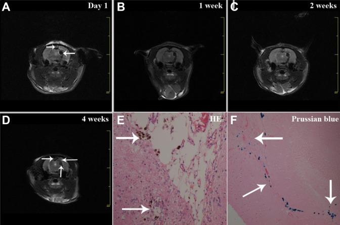
Figure 4.
Migration of implanted iPS cells-induced NSCs in TBI rat brains. T2*-weighted MRI scan was performed after iPS cells-induced NSCs (SPIOs-labeled) transplantation, showing pronounced hypointense signals at the cell injection site (A, as indicated by the lower white arrow). The dark signals gradually spread to the border of the damaged brain area from 1 (B) and 2 weeks (C) to 4 weeks (D, the left white arrow indicates the brain lesion, the lower white arrow indicates the cell injection site, the right white arrow indicates “migrated” signals). Hematoxylin-eosin staining (E) and Prussian blue staining of brain sections showed the presence of SPIOs-labeled NSCs with blue particles (F, the upper white arrow indicates NSCs in the brain lesion area, the lower white arrow indicates migrated NSCs, the right white arrow indicates the cell injection site).

