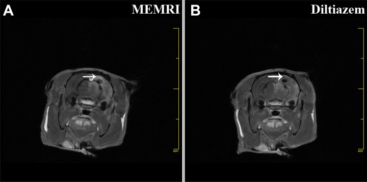
Figure 5.
ME-MRI tracking of iPS cells-induced NSCs function in TBI rats. TBI rats underwent ME-MRI scan, showing that a regional signal increase (A, white arrow) was produced in the lesion area. However, this enhancement could be blocked (B, white arrow) with diltiazem in another group of TBI rats, suggesting that localized neural activity was provided by implanted iPS cells-induced NSCs.

