Fig. 3.
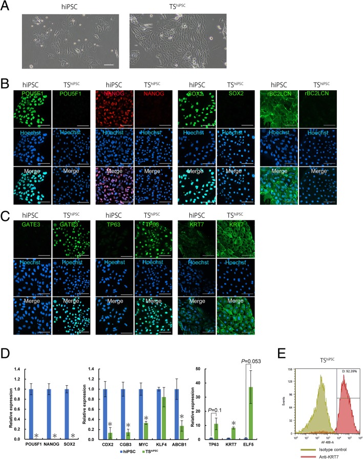
Morphology of and marker expression in hiPSCs and trophoblast cells derived from cysts (TShiPSC). a Phase-contrast images of hiPSCs and TShiPSC cells. Scale bar = 100 μm. b, c Bright-field and immunofluorescence images of hiPSCs and TShiPSC cells stained for POU5F1, NANOG, SOX2, and rBC2LCN (b) and GATA3, TP63, and KRT7 (c). Nuclei were stained with Hoechst 33342. Scale bar = 100 μm. d Analysis of pluripotency and trophoblast gene expression by qRT-PCR in hiPSCs and TShiPSC cells grown on laminin-coated dishes for 3 days. Expression levels were calculated relative to those of GAPDH and normalized to those of control hiPSCs. Values are the means ± SEMs (n = 4). Significance of differences was determined by Student’s t tests. *P < 0.05 versus the hiPSC group. e Flow cytometry histograms of KRT7 expression in TShiPSC cells. Similar results were obtained for three independent cell samples

