Fig. 5.
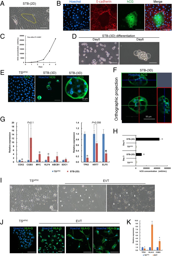
Directed differentiation of TShiPSC cells into STB- and EVT-like cells. a Phase-contrast image of TShiPSC cells containing multinucleated STB-(2D) cells (yellow-dotted line area). b Immunofluorescence images of TShiPSC cells containing multinucleated STB-(2D) cells stained for E-cadherin and hCG; nuclei were stained with Hoechst 33342. c Changes in hCG secretion by TShiPSC cells over 5 days. Data are presented as means ± SEMs (n = 4). Significance of differences was determined by one-way analysis of variance with repeated measures. d Phase-contrast images of STB-(3D) cells in low-adherence culture dishes on days 0 and 6. e Immunofluorescence images of TShiPSC cells and STB-(3D) cells stained for hCG. f Immunofluorescence images of STB-(3D) cells at z-position, showing the STB-(3D) cyst structure, which had several clear interior cavities and a thin enclosing wall. g Analysis of pluripotency and trophoblast gene expression by qRT-PCR in TShiPSC and STB-(3D) cells. Expression levels were calculated relative to those of GAPDH and normalized to those of TShiPSC cells. Values are the means ± SEMs (n = 4). Significance of differences was determined by Student’s t tests. *P < 0.05 versus the TShiPSC cell group. h Levels of hCG secreted by TShiPSC and STB-(3D) cells. For comparison, both TShiPSC and STB-(3D) cells were cultured in low-adherence Petri dishes. Data are presented as means ± SEMs (n = 3). i Phase-contrast images of TShiPSC and EVT cells. EVT cells had a mesenchyme-like morphology. The arrows indicate the presence of pseudopodia on EVT cells. j Immunofluorescence images of TShiPSC and EVT cells stained for HLA-G. k Analysis of TP63, HLA-G, and CGB gene expression by qRT-PCR in TShiPSC and EVT cells. Expression levels were calculated relative to those of GAPDH and normalized to those in TShiPSC cells. Values are the means ± SEMs (n = 4). Significance of differences was determined by Student’s t tests. *P < 0.05 versus the TShiPSC cell group. Unless otherwise noted, the scale bars in all phase-contrast and immunostaining images are 100 μm

