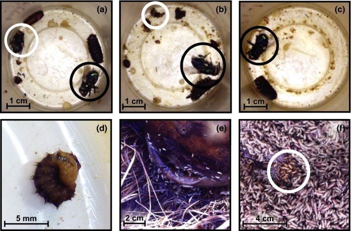
Figure 1.

Chrysomya rufifacies in the laboratory and in the field, including categorization of prey consumption level of Cochliomyia macellaria by C. rufifacies. Panels (a–c) are images representative of prey consumption level categorization. Black circles indicate C. rufifacies, and white circles indicate Co. macellaria. (a) No consumption—two pupal casings (one C. rufifacies and one Co. macellaria). (b) Partial consumption—one pupal casing (C. rufifacies) and part of prey remaining. (c) Total consumption—one pupal casing (C. rufifacies) and no evidence of prey remaining. (d) When C. rufifacies is predating, it will wrap itself around the body of its prey item. (e) Spatial segregation between C. rufifacies and C. macellaria is frequently observed on human remains in the field. Chrysomya rufifacies is general found at the interface between the donation and the soil, with Cochliomyia macellaria on the surface (not pictured). (f) On the rare occasions that both species occupy the same area, the two species are found in separate masses (C. rufifacies in white circle)
