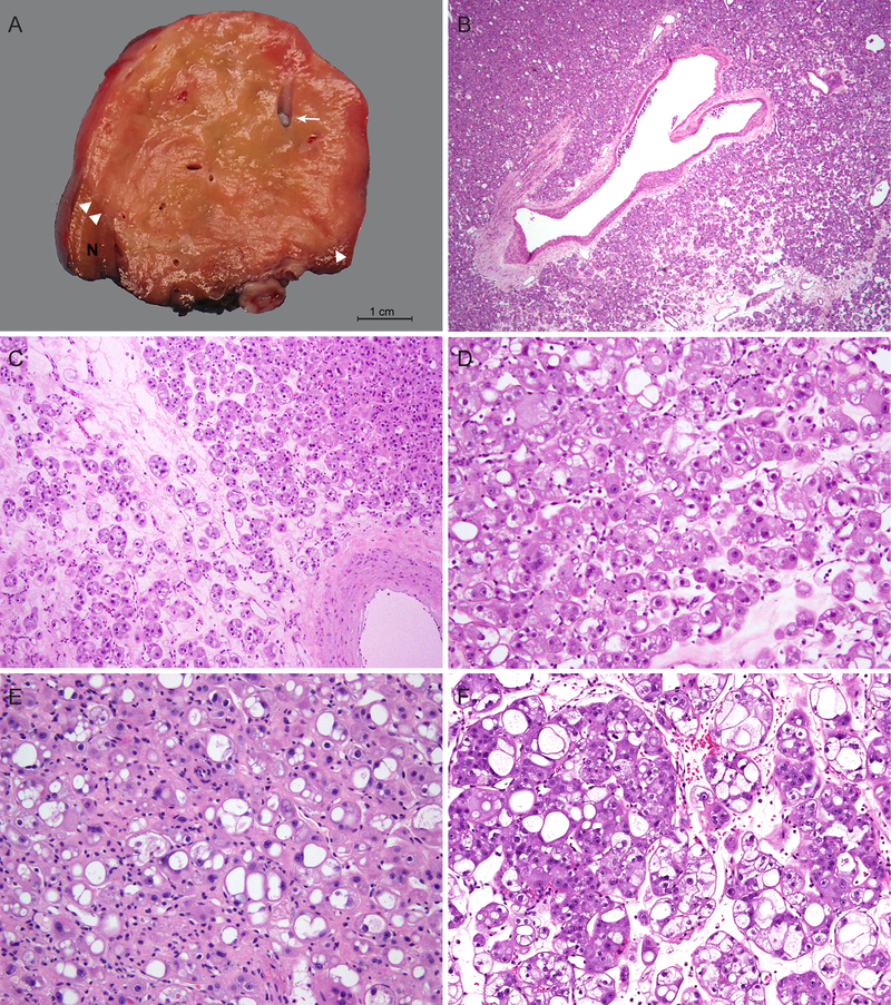
Figure 1.
Macroscopic and microscopic features of a distinct group of RCC with eosinophilic and vacuolated cytoplasm. (A) Tumors are well-circumscribed with tan-yellow to tan-brown, mostly solid cut surfaces. Arrow marks intratumoral vessels with medium to large caliber; arrowheads mark the boundary between tumor and adjacent renal parenchyma. (B-C) Tumors consist of nests of eosinophilic cells in a hypocellular and often edematous stroma. Dispersed single or minute clusters of cells are also present. Note an absence of foamy histiocytes or lymphocytic infiltrates in the stroma. (D-F) Tumor cells show round nuclei with conspicuous to prominent nucleoli and eosinophilic, granular and vacuolated cytoplasm. Vacuolization varies from numerous small intracytoplasmic vesicles to large spaces almost occupying the entire cytoplasm. (D) Case 1, (E) Case 4, (F) Case 6.

