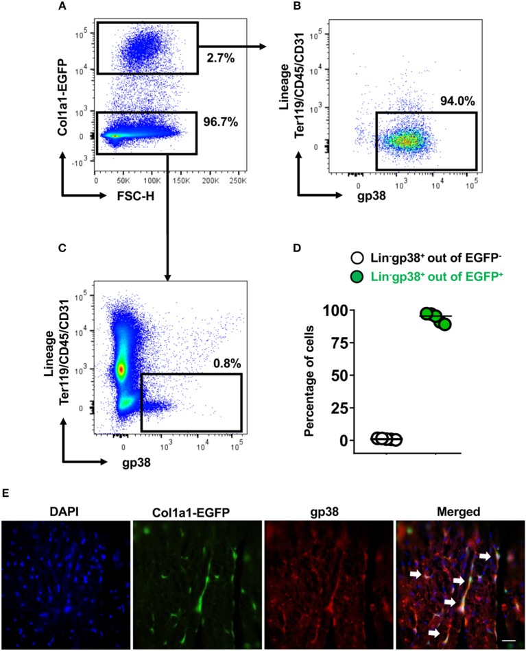
Figure 3.
Cardiac Lin−gp38+ cells represent collagen I producing cells. Representative flow cytometry analysis of cells isolated from hearts of Col1a1-EGFP tg mice. Cellular debris below 50 k in FSC-A were excluded in SSC-A/FSC-A plots. Live cells were first gated as Col1a1-EGFP+ or Col1a1-EGFP− (A) and then analyzed for expression of Lin and gp38. The percentage of Lin−gp38+ cells was quantified from Col1a1-EGFP+ cells (B) and Col1α1-EGFP− cells (C). (D) Shows quantification of Lin−gp38+ cell percentages from EGFP− (white circles) and EGFP+ (green circles) cells (n = 5). (E) Shows representative staining of gp38 (red) and EGFP expression (green) indicating the collagen I-producing cells in the cryosections from hearts of Col1a1-EGFP tg mouse. DAPI stains cell nuclei. Bar = 50 μm.

