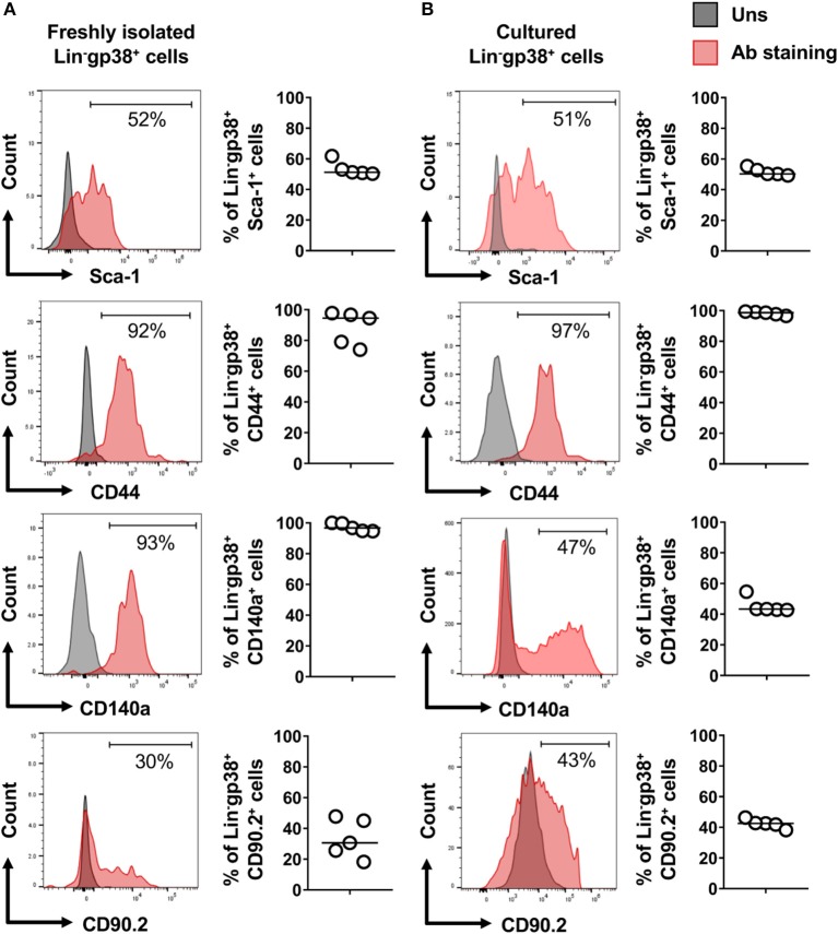
Figure 4.
Expression of mesenchymal cell markers on cardiac Lin−gp38+ cells. Representative flow cytometry histograms (data are representative for 1 out of 5 independent experiments; gray color indicates unstained controls) and quantification of the indicated mesenchymal markers on Lin−gp38+ cells of single cell suspension obtained from mouse heart (A) and cardiac Lin−gp38+ cells expanded in vitro for 3 passages (B) (n = 5).

