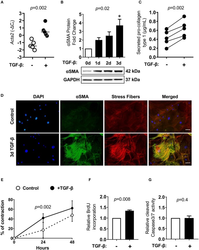
Figure 5.
Cardiac Lin−gp38+ cells response to TGF-β1 stimulation. Cardiac Lin−gp38+ cells were isolated from hearts of C57BL/6 mice as described in Figure 1 and expanded in vitro. (A) Shows Acta2 mRNA expression in cardiac Lin−gp38+ cells cultured in the presence or absence of TGF-β1 (10 ng/mL) for 24 h (n = 5, p-value calculated with Mann-Whitney U-test). (B) Shows quantification and representative immunoblots for αSMA protein levels in lysates of Lin−gp38+ cells cultured in the presence or absence of TGF-β1 (10 ng/mL) for the indicated time [n = 5, p-values computed using Kruskal-Wallis test followed by the Dunn's multiple comparisons test, *p < 0.05 (post-hoc test vs. control)]. In (C), results from ELISA for pro-collagen type 1 are presented. Lin−gp38+ cells were seeded at passage 3 and treated with or without TGF-β1 (10 ng/mL) for 7 days. Supernatants were used to assess the amount of pro-collagen type 1 secreted from Lin−gp38+ cells (n = 5, p-value calculated with paired t-test). (D) Shows representative staining of αSMA (green) and phalloidin (red) in Lin−gp38+ cells cultivated in the presence or absence of TGF-β1 (10 ng/mL) for 3 days. DAPI stains cell nuclei. Bar = 50 μm. (E) Shows quantification of Lin−gp38+ cells contractility precultured in the presence or absence of TGF-β1 (10 ng/mL) for 3 days (n = 6, p-value calculated with Sidak's multiple comparison test). (F) Indicates quantification of BrdU incorporation by Lin−gp38+ cells cultured in the presence or absence of TGF-β1 (10 ng/mL) for 24 h (n = 5, p-value calculated with Mann-Whitney U-test). (G) Shows quantification of cleaved caspase 3/7 activity in Lin−gp38+ cells cultured in the presence or absence of TGF-β1 (10 ng/mL) for 24 h (n = 4, p-value calculated with Mann-Whitney U-test).

