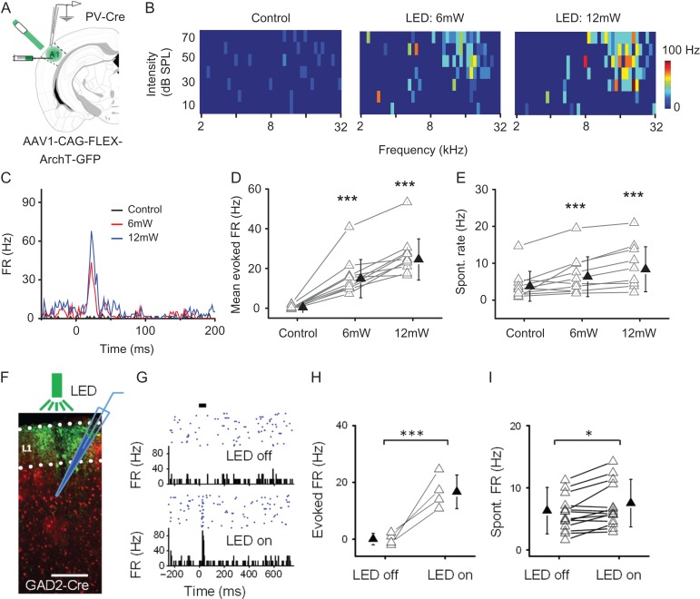
Figure 6.
Manipulations of the sparse level by 2 methods. I. Suppressing PV inhibitory neurons reduces sparseness level. (A) Injection of AAV encoding Cre-dependent ArchT into A1 of PV-Cre mice. Green LED light was applied to A1 surface. Loose-patch recording was made in A1. (B) Mapped TRFs in a putative L2/3 excitatory neuron in the control condition (left) and when tone stimuli were coupled with LED stimulation at 6 mW (middle) and 12 mW (right) power. (C) PSTH for spike responses to all test tones for the same cell shown in (B). (D) Average firing rate evoked by the best tone as observed in the condition of 6- and 12-mW LED stimulation. N = 10 cells. ***P < 0.001, one-way ANOVA and post hoc test (compared with control). (E) Spontaneous firing rate measured within 50-ms window preceding tone onset in control and LED stimulation conditions. ***P < 0.001, one-way ANOVA and post hoc test (compared with control). II. Suppressing L1 inhibitory neurons reduces sparseness level. (F) Expression of ArchT (green) in GAD2-Cre::Ai14 mice. Green LED was applied to A1 surface. Loose-patch recording was made from L2/3. (G) Responses of a putative L2/3 excitatory neuron to noise stimulation (thick bar) in the control condition (top) and when noise was coupled with LED illumination (bottom). (H) Noise-evoked firing rates of 4 L2/3 neurons converted from NR to R cells. ***P < 0.001, t-test. (I) Spontaneous firing rates of 15 L2/3 neurons examined in the control and LED on conditions. *P < 0.05, t-test.

