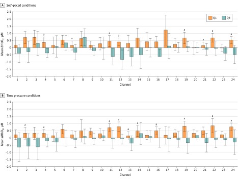
Figure 2. Channel Activation in Quartile 1 (Q1) and Quartile 4 (Q4) Residents.
Between-group channelwise differences in prefrontal change in oxygenated hemoglobin concentration (ΔHbO2) during self-paced (A) and time pressure (B) conditions. The Q1 residents demonstrate significantly greater activation compared with Q4 residents in 8 channels in the bilateral ventrolateral prefrontal cortex in the self-paced condition and in 10 channels in the bilateral ventrolateral prefrontal cortex and left dorsomedial prefrontal cortex in the time pressure condition. Error bars represent the 95% confidence interval.
aP < .05 (independent-samples t test).

