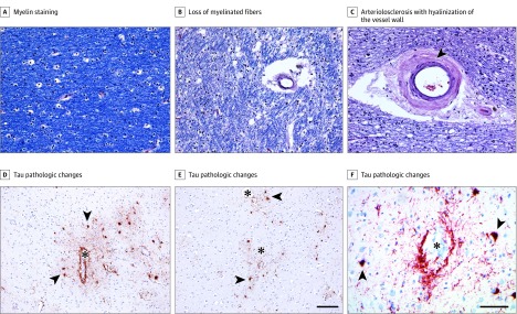
Figure 2. White Matter Rarefaction, Arteriolosclerosis, and Dorsolateral Frontal Cortex Tau Pathologic Changes in Participants With Chronic Traumatic Encephalopathy.
A, Luxol fast blue with hemotoxylin-eosin histochemical staining shows robust myelin staining (blue) in a former US football college player in his early 40s who was neuropathologically diagnosed with chronic traumatic encephalopathy (CTE) (stage I/II) who was not determined to have had antemortem dementia. B and C, In a man who had played professional US football, was in his mid-80s, had been neuropathologically diagnosed with CTE (stage III/IV), and was determined by consensus to have dementia, there was severe loss (3+) of myelinated fibers (B), as well as marked arteriolosclerosis with hyalinization of the vessel wall (C, arrowhead). D-F, Immunohistochemical staining for tau pathologic changes (antibody AT8) in the dorsolateral frontal cortex shows perivascular accumulations of abnormal tau at the sulcal depths within neurons and cell processes (asterisks indicate the lumen of cortical vessels; arrowheads, examples of pretangles and tangles within neurons). The scale bar for A-C and F is 100 μm; for D and E, 150 μm.

