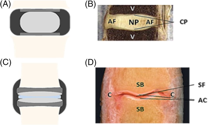
Figure 1.

A and B, Anatomy of the human intervertebral disc and articular joint.29, 30 A, The intervertebral disc consists of a nucleus pulposus (light gray), surrounded by an annulus fibrosus (black), and is between two cartilaginous endplates (dark gray) that adhere to the adjacent vertebrae (beige). B, An image of a sagittal section of an intervertebral disc. NP: nucleus pulposus; AF: annulus fibrosus; CP: cartilaginous endplates; V: vertebrae. C, The articular joint consists of articular cartilage (light gray), that lies over the subchondral bone (dark gray) of the adjacent joints, and is divided by synovial fluid (light blue). The capsule (black) surrounds the articular joint. D, An image of a sagittal section of an articular joint. AC: articular cartilage; C: capsule; SB: subchondral bone; SF: synovial fluid
