Figure 3.

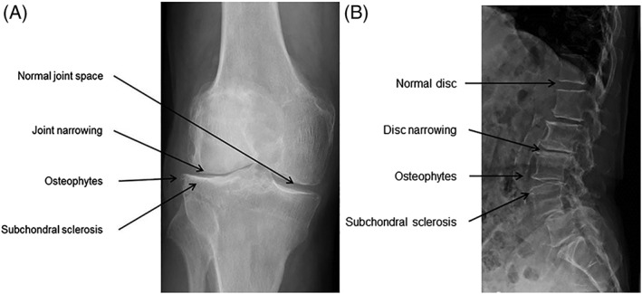
A and B, Radiological examples in the degenerated articular joint and intervertebral disc. Both OA (A) and DD (B) are radiologically characterized by loss of joint space, the formation of osteophytes and subchondral sclerosis

A and B, Radiological examples in the degenerated articular joint and intervertebral disc. Both OA (A) and DD (B) are radiologically characterized by loss of joint space, the formation of osteophytes and subchondral sclerosis