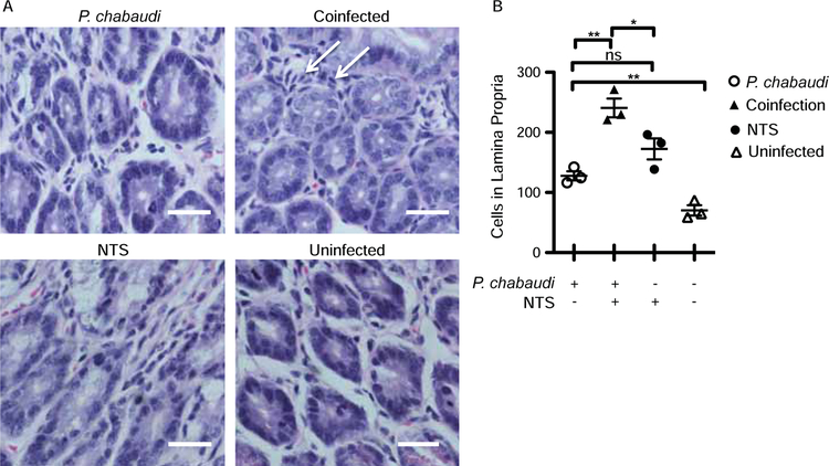
Figure 3.
Malarial parasite-infected animals showed increased cellularity within the lamina propria. C57Bl/6 mice were infected with P. chabaudi, and co-infected mice received Salmonella Typhimurium gavage 4 days post malaria infection and were sacrificed 11 days post malarial infection (expt.2). Sections representing the jejunum of the small intestine were stained with H&E. (A) Representative image (40X) showing a cross-section of the lamina propria in the jejunum. Arrows indicate cellular infiltration. The scale bar represents 25mm. (B) Quantification of the number of cells in the lamina propria of sections of the jejunum. Statistically significant differences between groups are indicated with *p ≤ 0.05 and **p ≤ 0.01.

