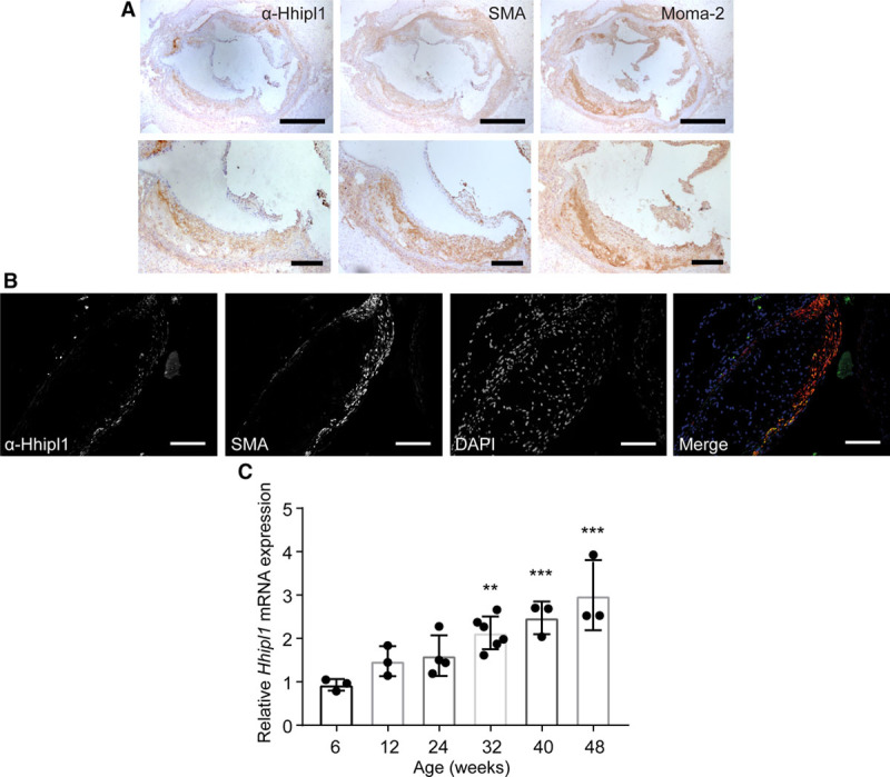
Figure 3.

Hhipl1 expression in atherosclerotic plaques. A, Representative immunohistochemical staining with anti–α smooth muscle actin antibody (SMA), anti-Hhipl1, and MOMA-2 in aortic root lesions from 18-week-old Apoe−/− mice fed Western diet for 12 weeks. Bars, 500 μm (top) and 200 μm (bottom). B, Immunofluorescent staining of aortic root lesion with DAPI, SMA, and anti-Hhipl1. Bars, 100 μm. C, Hhipl1 mRNA expression relative to Rpl4 in the aortic arch of 6- to 48-week-old Apoe−/− mice. n=3–6 mice per time point. Error bars represent mean±SD. HHIPL1 indicates hedgehog interacting protein-like 1. *Post hoc comparisons with 6-week time point. **P≤0.01, ***P≤0.001.
