Figure 4.

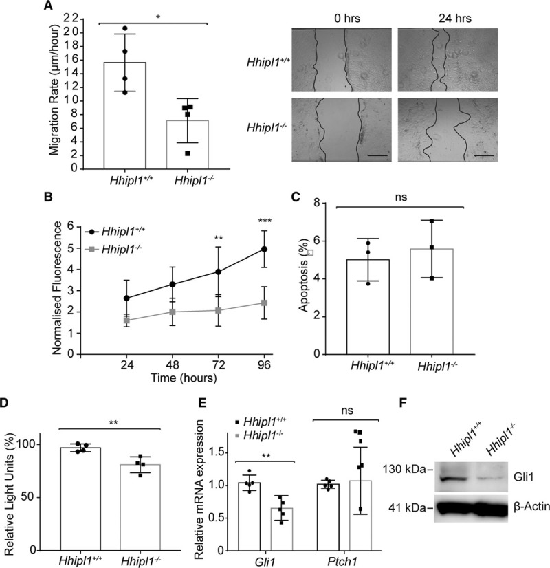
HHIPL1 regulates mouse AoSMC migration and proliferation. A,Migration rate of Hhipl1−/− and wild-type AoSMCs in a scratch wound assay over a period of 24 hours (n=4). Representative images are shown (right). B, Proliferation of Hhipl1−/− and wild-type AoSMCs over a period of 96 hours (n=4). *Significant post hoc comparisons at 72 and 96 hours. C, Proportion of apoptotic wild-type and Hhipl1−/− AoSMCs. D, Gli-luciferase activity in SHH-LIGHT2 cells co-cultured with either wild-type or Hhipl1−/− AoSMCs; n=4. E, Gli1 and Ptch1 mRNA expression relative to Rplp0 in wild-type and Hhipl1−/− AoSMCs; n=5. Error bars represent mean±SD. *P≤0.05, **P≤0.01, ***P≤0.001. F, Western blot showing Gli1 expression in wild-type and knockout cells. β-actin was used as a loading control. AoSMC indicates aortic smooth muscle cell; HHIPL1, hedgehog interacting protein-like 1; and SHH, sonic hedgehog.
