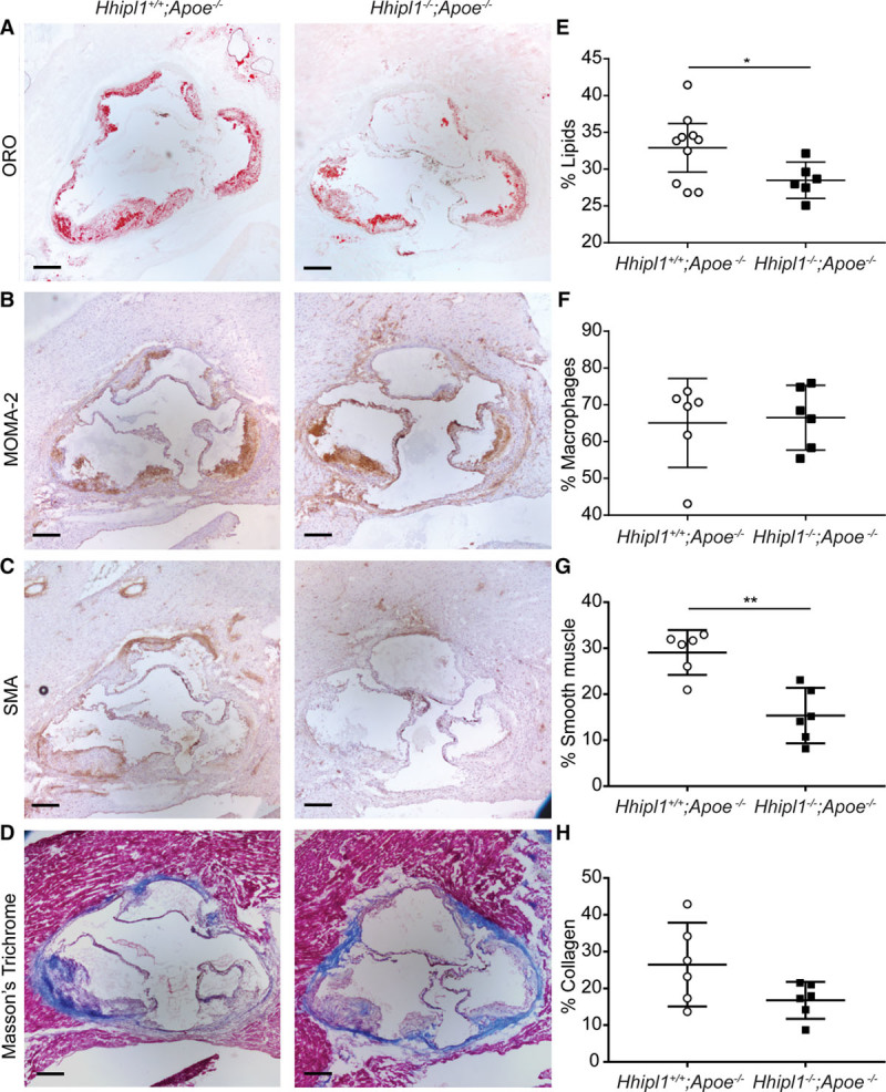
Figure 7.

Hhipl1 deficiency reduces smooth muscle cell content in Hhipl1−/−;Apoe−/−atherosclerotic lesions. Representative photomicrographs of atherosclerotic lesion components in Hhipl1+/+;Apoe−/− and Hhipl1−/−;Apoe−/− mice. A, Oil red O (ORO) staining for lipids; C, MOMA-2 staining for macrophages; E, anti-α smooth muscle actin (SMA) staining for smooth muscle cells; and G, Masson’s trichrome for collagen. Bars, 200 μm. Percentage coverage (average of 9 sections per animal) of (B) lipids, (D) macrophages, (F) smooth muscle cells (P=0.001), and (H) collagen (n=6–10 per group). Error bars represent mean±CI. HHIPL1 indicates hedgehog interacting protein-like 1. *P≤0.05, **P≤0.01. Bar, 200 μm.
