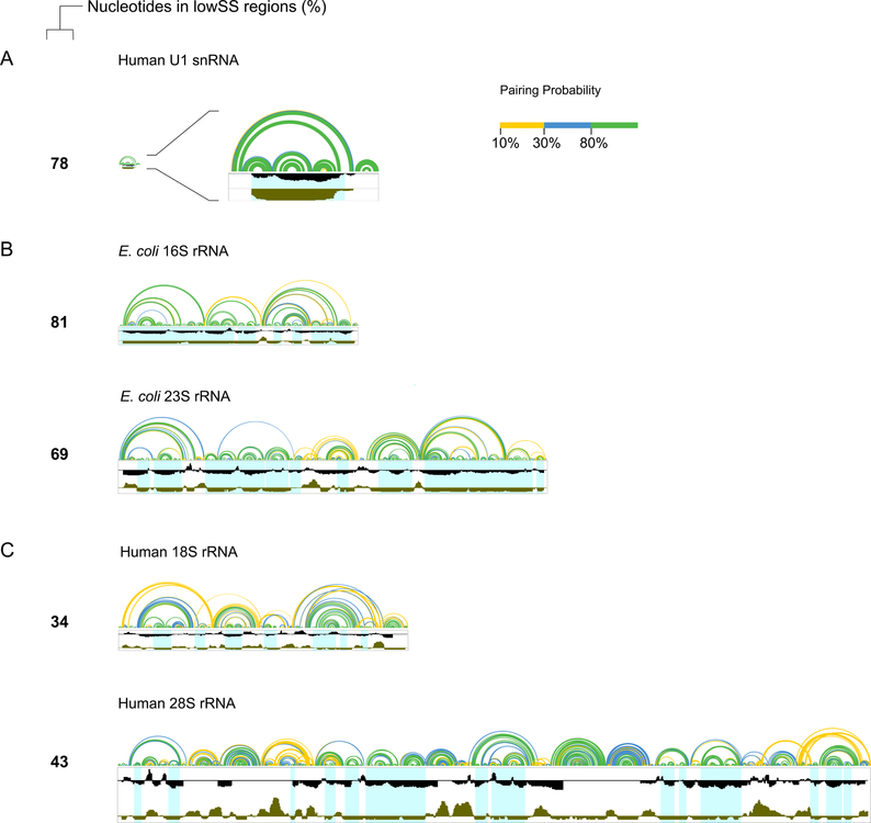
Figure 5. Extent of well-determined structures in small and large RNAs.
(A) human U1 snRNA, (B) E. coli 16S and 23S rRNA, and (C) human 18S and 28S rRNA. Arcs represent modeled base pairing probabilities (colors defined in key). Windowed SHAPE reactivities and entropy are shown in black and brown, respectively. Well-structured regions are highlighted in light blue. Percentages of nucleotides in lowSS regions exclude positions with no SHAPE data (primarily located near the 5’ and 3’ ends of each RNA, and one central section of the 28S rRNA).

