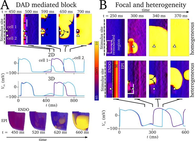
Fig 10. Mechanisms of SCRE mediated conduction block.
A–Demonstration of DAD-mediated conduction block in 2D (upper panels) and 3D (lower panels). In both cases, two stimuli were applied to one side of the tissue (left edge of the 2D sheet; ENDO surface in 3D) at a coupling interval of 500 ms, with SCRE induced DADs interrupting the second applied stimulus. Spatial snapshots cover the time just before and during this second stimulus. The locations of the cells from which the AP traces are taken are indicated by the triangular markers in the 2D sheets; for the 3D case, the traces correspond to a region which did (blue) and did not (purple) exhibit conduction block. Solid white lines represent sites of conduction block. B–Demonstration of spontaneous focal excitation leading to different behaviour in electrically homogeneous or heterogeneous tissue. The purple trace corresponds to the homogeneous condition, in which the focal excitation propagates uniformly; the blue trace corresponds to the heterogeneous condition in which focal excitation propagates non-uniformly following conduction block. The triangular marker indicates the site from which the AP traces were extracted, and the region of reduced IK1 is highlighted by the dashed-white rectangle. The stimulus is applied once to the left edge (ENDO region) of the tissue at t = 0 ms; the second excitation is spontaneously induced.

