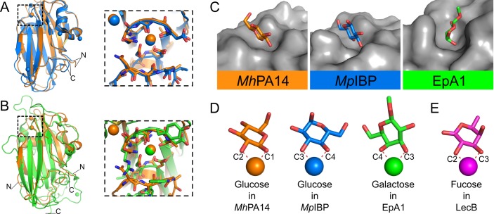
Fig 7. Structural comparison of MhPA14 to other PA14 domains.
Structural alignment of MhPA14 (orange) to either A) MpIBP PA14 (blue) or B) EpA1 A domain (green). Close-up views of the sugar-binding site for both alignments are shown in boxes. C) Sugar-binding pockets of these three PA14 structures. Protein topology is coloured grey, sugars are coloured with oxygen in red, nitrogen in blue, and the carbon atoms in the respective colours of their structures shown in A and B. D) The orientation of these same sugars as they coordinate to the calcium ions in their respective structures. E) Orientation of fucose (purple) from LecB.

