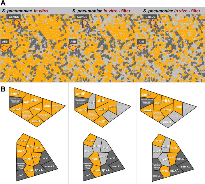
Fig 2. Identification of ComDE and AliB in S. pneumoniae D39 from in vitro culture or in vivo infection samples.
(A) Voronoi Treemaps of predicted and detected pneumococcal proteins. S. pneumoniae D39 was cultured under various in vitro conditions, and a map of in vitro identified proteins was generated (left panel). Pneumococcal proteins identified in control reactions with a trypsin-digestion of pneumococci on a filter (middle panel). These pneumococci represent bacteria from a culture that was also used to infect mice. Proteins identified in pneumococci recovered from the CSF of mice (n = 5 samples each of 4 mice), enriched by sequential centrifugation on a filter, and digested by trypsin (right panel). Gray spheroids represent annotated protein entries from the SEED with light gray not identified in sample and dark gray never identified. Orange spheroids represent identified proteins by MS. (B) Identified proteins are depicted in enlarged regions of Voronoi treemaps. ComD, ComE (upper left), and AliB (lower left) were not identified in S. pneumoniae D39 from in vitro culture samples, while all three proteins were identified in samples from in vivo infection (upper and lower right).

